
🫁 The Lung’s Quiet Defense System
 ToothOps3 min read

How the Lymphatic Network Shapes Immunity — and What TB Reveals About Disease Patterns
When clinicians interpret lung disease, they often start with airspaces and blood vessels.But many of the most telling clues travel a quieter route.
The pulmonary lymphatic system regulates fluid balance, filters pathogens, and coordinates immune decisions.Understanding how it works explains why diseases like tuberculosis (TB) behave the way they do — on imaging, clinically, and over time.
🧠 ToothOps Mental Model:
Drainage → Detection → Decision
This framework organizes everything that follows.
- Drainage → prevents pulmonary edema and preserves gas exchange
- Detection → samples antigens and activates immune cells
- Decision → immune system escalates, contains, or tolerates
Most lymphatic findings represent where the system is in this sequence.
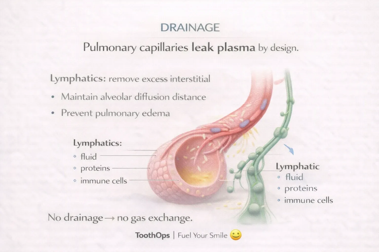
1️⃣ Drainage: Why the Lung Depends on Lymphatics
Pulmonary capillaries constantly leak plasma into interstitial tissue.Lymphatic capillaries — thin, blind-ended vessels — collect this fluid before it interferes with oxygen exchange.
Key roles:
- Maintain dry alveoli
- Regulate interstitial pressure
- Prevent pleural fluid accumulation
🧠 Clinical insight: Pulmonary edema reflects lymphatic overload, not lymphatic absence.
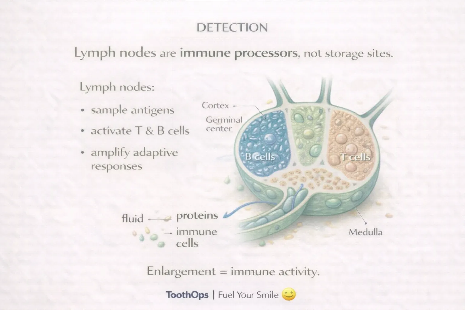
2️⃣ Detection: Lymph Nodes as Immune Sensors
Lymph nodes are not passive filters. They are active immune processors.
They enlarge when:
- Antigens are presented
- Lymphocytes clonally expand
- Macrophages accumulate pathogens
Tender, mobile nodes → rapid immune activationFirm, persistent nodes → chronic stimulation or infiltration
💡 Enlargement often signals immune work, not disease spread.
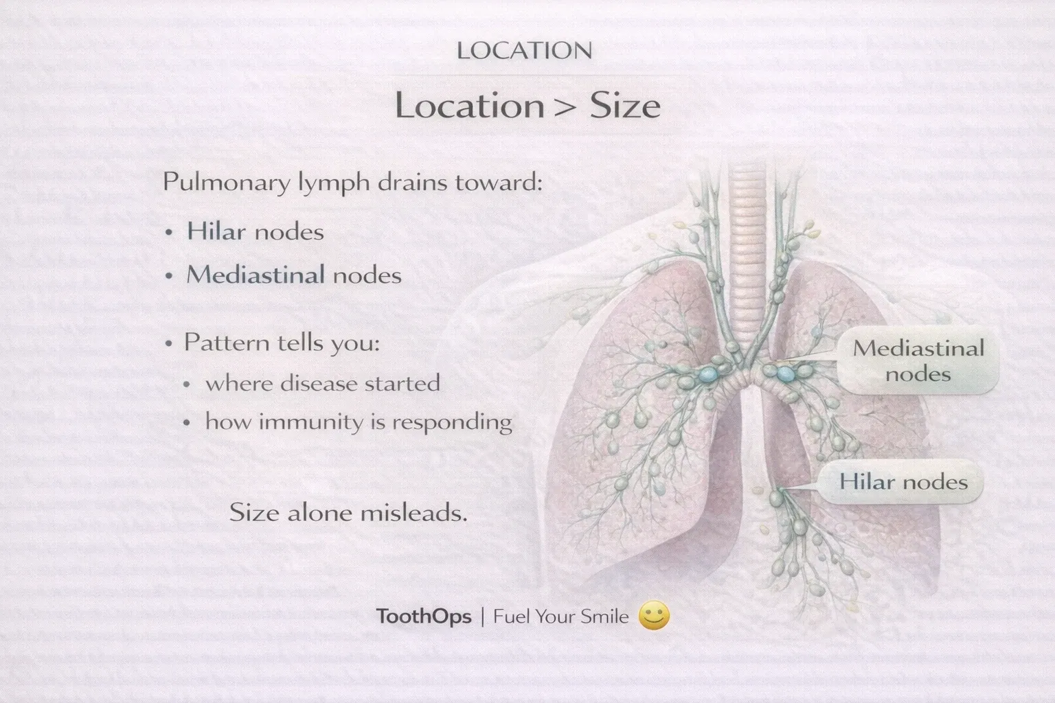
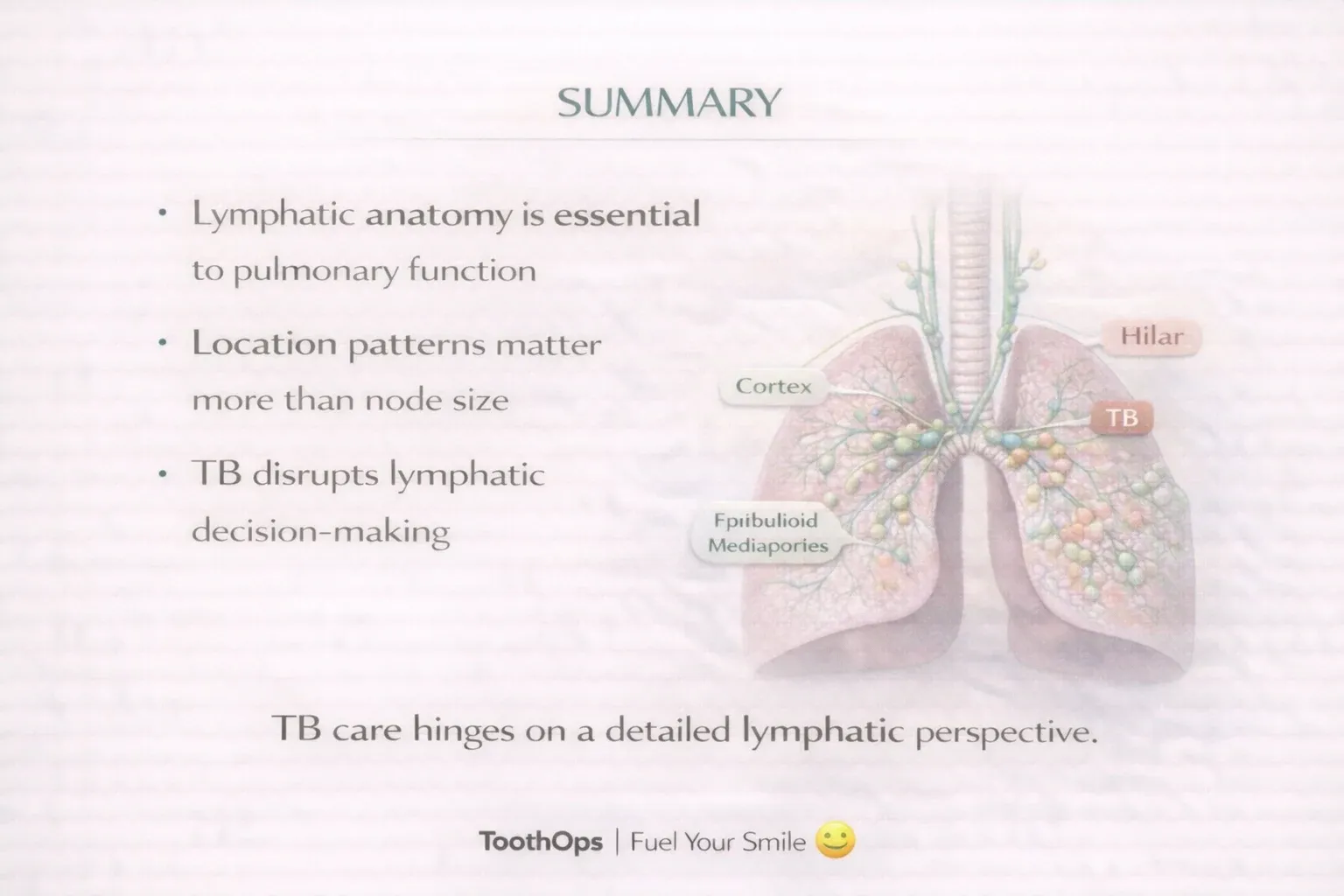
3️⃣ Why Location Matters More Than Size
Pulmonary lymph drains toward:
- Hilar nodes (central lung drainage)
- Mediastinal nodes (final thoracic checkpoints)
Patterns matter:
- Symmetric bilateral nodes → immune-mediated disease
- Asymmetric focal nodes → localized pathology
🧠 Node location often narrows the differential faster than node size.
4️⃣ Cellular Architecture: Why Granulomas Form
Inside lymph nodes:
- Cortex → B-cell antibody responses
- Paracortex → T-cell coordination
- Medulla → macrophage filtration
When pathogens resist clearance, immune cells organize into granulomas — structured containment units designed to limit spread.
Granulomas are not failure. They are controlled stalemates.
5️⃣ TB as a Lymphatic Disease (Not Just Pulmonary)
TB engages the lymphatic system early.
Classic features:
- Lung lesion + draining lymphadenopathy
- Formation of the Ghon complex
- Early containment without complete eradication
Primary TB often involves lymph nodes prominently, especially in younger patients.
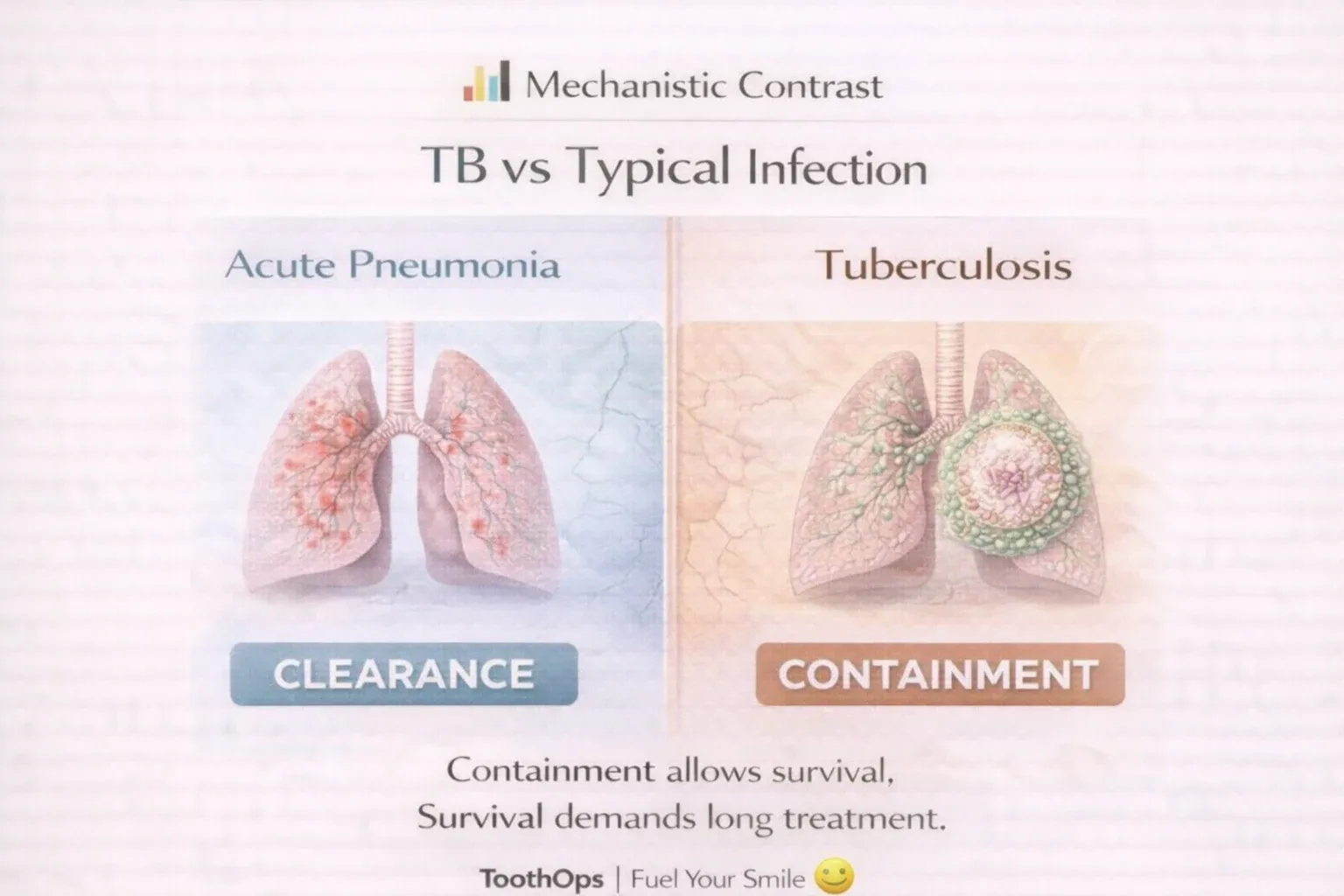
📊 TB vs Typical Pulmonary Infections
| Feature | Typical Pneumonia | Tuberculosis |
|---|---|---|
| Onset | Acute | Subacute / chronic |
| Immune goal | Clearance | Containment |
| Lymph nodes | Transient | Persistent |
| Histology | Neutrophils | Granulomas |
| Treatment | Short | Prolonged |
🧠 TB survives because containment can last years.
⚠️ Clinical Misinterpretation Trap
Enlarged mediastinal lymph nodes ≠ advanced TB.
In TB, lymphadenopathy often reflects:
- Early immune containment
- Active antigen processing
- Structured immune surveillance
Mislabeling this as “late disease” can delay diagnosis or misguide management.
6️⃣ Why TB Requires Time (Biology Explains Treatment)
Granulomas limit bacterial spread — but don’t eliminate TB.
Within granulomas:
- Bacteria enter low-metabolic states
- Antibiotics penetrate slowly
- Immune clearance plateaus
This explains:
- Cavitation
- Latency
- Need for prolonged therapy
Containment ≠ cure.
7️⃣ Reading Imaging Through Lymphatic Logic
When evaluating scans, ask:
- Where does lymph from this region drain?
- Is the immune system containing or failing?
- Does oxygen tension explain lesion location?
This transforms images into mechanistic narratives, not isolated findings.
8️⃣ Transfer Learning: Why This Matters Beyond TB
This same lymphatic reasoning applies to:
- Sarcoidosis
- Lymphoma
- Metastatic spread
Disease patterns follow drainage pathways, not randomness.
🌱 Final ToothOps Takeaway
The lymphatic system doesn’t announce itself — but it shapes disease behavior.
TB teaches us:
- Containment can look like stability
- Nodes tell stories
- Biology sets the pace
Understanding systems builds confidence.
@ToothOps | Fuel Your Smile 😊 Stay tuned for more insights and educational content in our blog.
Disclaimer: Content is for educational purposes only and not a substitute for medical or dental care.
© 2025 ToothOps | All Rights Reserved.