
🫁 TB vs Pneumonia Made Simple: How X-Rays & Exam Signs Finally Make Sense
 ToothOps3 min read
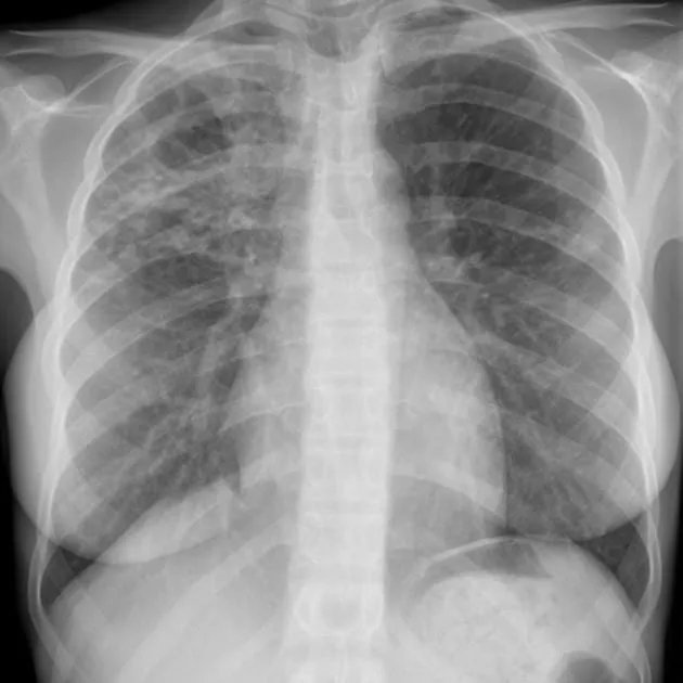
If chest X-rays and lung exam terms like egophony or apical crackles feel confusing, you’re not alone.Most students try to memorize them — when what you really need is a mental model.
This post walks you through TB vs pneumonia the ToothOps way:calm, mechanism-first, and confidence-building.
1️⃣ Start With One Question (This Solves 80%)
What is happening to the lung tissue?
🫁 Pneumonia → the lung is filled🦠 Tuberculosis (TB) → the lung is being destroyed
🧠 Analogy Box
- Pneumonia = a sponge soaked with water
- TB = wood slowly eaten by termites
Everything you see on X-ray and physical exam flows from this difference.
2️⃣ Chest X-Ray: What You’re Actually Looking For
🫁 Pneumonia on X-Ray
Mechanism Acute inflammation fills alveoli with fluid, pus, and immune cells.
What you see
- Consolidation (white, solid-looking lung)
- Often limited to one lobe
- Air bronchograms (black branching airways inside white lung)
- Changes appear quickly (days)
- Improves with antibiotics
🧠 Why air bronchograms appearAlveoli are fluid-filled (white), but bronchi still contain air (black).That contrast is classic for pneumonia.
🦠 TB on X-Ray
MechanismChronic immune response forms granulomas → tissue breakdown → cavities.
What you see
- Upper-lobe disease
- Cavitary lesions (holes)
- Patchy, irregular opacities
- Fibrosis or volume loss over time
- Changes develop slowly (weeks–months)
🧠 **Why upper lobes?**TB thrives in high-oxygen environments, which the upper lobes provide.
📊 One-Look X-Ray Comparison
| Feature | Pneumonia | TB |
|---|---|---|
| Core process | Alveoli filled | Tissue destroyed |
| Key word | Consolidation | Cavitation |
| Location | Any lobe | Upper lobes |
| Speed | Fast | Slow |
| Reversibility | Yes | Often scars |
3️⃣ Physical Exam: What Your Hands & Ears Are Detecting
🔊 Crackles (Rales)
What crackles meanAir popping open fluid-filled airways.
-
Pneumonia:
- Localized crackles
- Appear early
- Loud over affected lobe
-
TB:
- Often apical crackles
- Subtle or absent early
- Appear late
🧠 Fluid makes noise. Holes don’t.
🗣️ Egophony (The “EEE → AAA” Sign)
What it isPatient says “EEE,” examiner hears “AAA.”
Why it happensSolid tissue transmits sound better than air.
- Pneumonia:✅ Egophony present (alveoli are solid with fluid)
- TB:❌ Usually absent (cavities disrupt sound)
💡 Pro Tip Egophony = consolidation → think pneumonia.
🥁 Percussion (Chest Tapping)
What you’re testingHow much air is underneath.
| Sound | Meaning |
|---|---|
| Resonant | Air |
| Dull | Fluid or solid |
-
Pneumonia:
- Dull to percussion (fluid replaces air)
-
TB:
- Often normal early
- May become dull later with fibrosis
- Mixed sounds near cavities
🧠 Early dullness strongly favors pneumonia.
4️⃣ Chest Pain: Pleuritic vs Non-Pleuritic
🔪 Pleuritic Chest Pain
Sharp pain, worse with breathing or coughing.
- Pneumonia:✅ CommonAcute inflammation irritates the pleura
- TB:❌ Usually absent earlyAppears only if pleura involved late
🧠 Acute inflammation hurts. Chronic destruction is quieter.
5️⃣ Put It All Together (This Is the Framework)
🫁 Pneumonia
- Fluid-filled lung
- Loud exam findings
- Obvious symptoms
- Fast timeline
- Reversible
🦠 TB
- Destroyed lung
- Quiet early exam
- Chronic, vague symptoms
- Slow timeline
- Often leaves scars
🧠 Final One-Look Summary
| Feature | Pneumonia | TB |
|---|---|---|
| X-ray | White consolidation | Upper-lobe cavities |
| Crackles | Loud, early | Apical, late |
| Egophony | Yes | No |
| Percussion | Dull early | Often normal early |
| Chest pain | Pleuritic | Mild or absent |
| Timeline | Days–weeks | Weeks–months |
🌱 Confidence Takeaway
You don’t need to memorize lung signs.
Just ask:
Is the lung filled — or being destroyed?
Once you answer that, the X-ray and exam findings make sense naturally.
@ToothOps | Fuel Your Smile 😊
Stay tuned for more insights and educational content in our blog.
Disclaimer: Content is for educational purposes only and not a substitute for medical or dental care.
© 2025 ToothOps | All Rights Reserved