
🦷 Why the Whites of the Eyes Turn Blue
 ToothOps6 min read
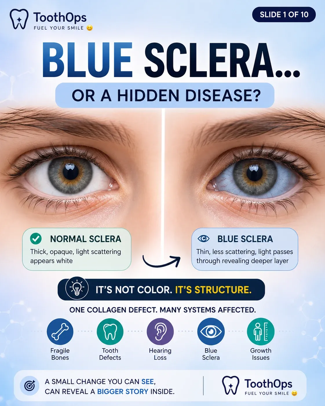
How Collagen Failure Changes Light, Structure, and What We See
🧠 Start Here: The Real Question
When clinicians see blue sclera, many think:
“That’s just an eye finding.”
But the real question is:
👉 Why did the tissue become transparent enough to change color?
Because blue sclera is not simply a color problem.
It is:
- a structural problem
- a collagen problem
- a light-transmission problem
- and often a clue to systemic disease
⚙️ Core Rule
What we see in biology is often determined not by pigment—but by how light interacts with structure.
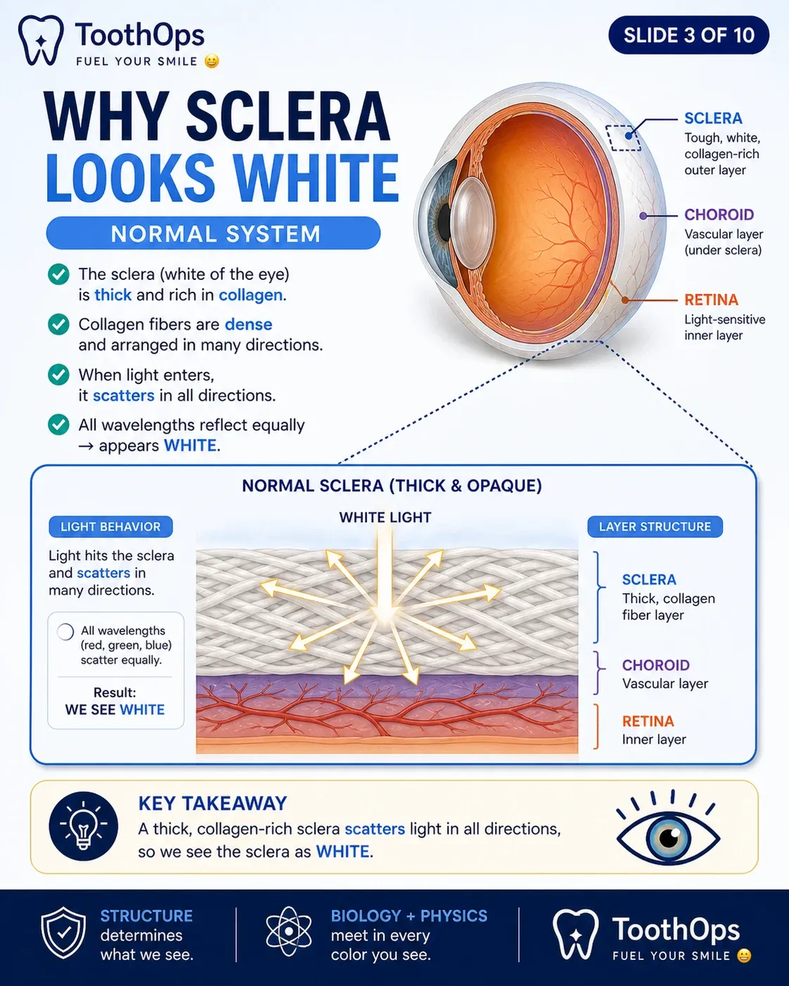
🪶 Big Picture: Why the Eye Normally Looks White
The sclera (“white of the eye”) appears white because it is:
- thick
- collagen-rich
- highly organized to scatter light
Normal sclera:
- reflects and scatters incoming light diffusely
- prevents deeper structures from being visible
👉 this creates the normal white appearance
🔬 The Molecular Foundation: Type I Collagen
Type I collagen is the major structural protein in:
- bone
- dentin
- ligaments
- sclera
🧬 Normal Collagen Structure
Healthy collagen contains:
- tightly packed triple helices
- glycine every third amino acid
- strong fibril cross-linking
This organization creates:
- tensile strength
- flexibility
- optical opacity
🧬 What Happens in Osteogenesis Imperfecta (OI)
In this case:
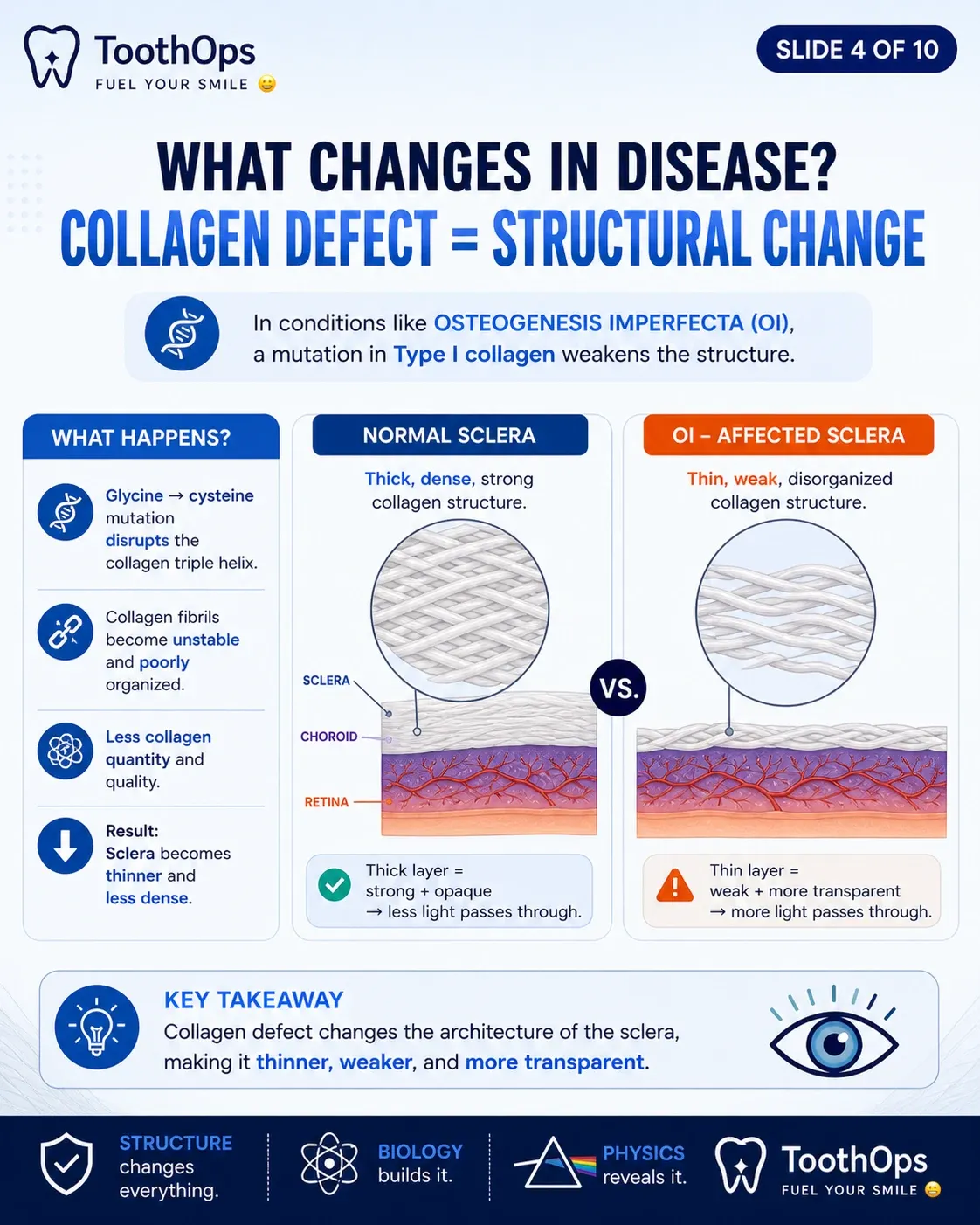
- glycine → cysteine mutation disrupts collagen helix formation
This leads to:
- abnormal collagen folding
- weak fibril assembly
- reduced structural integrity
🧠 Critical Insight
The same collagen defect weakening bone and dentin is also changing how light moves through the eye.
🔬 Tissue-Level Changes in the Sclera
The sclera is composed primarily of dense collagen bundles.
Normal sclera:
- thick collagen lamellae
- strong structural organization
- high light scattering
In OI:
- collagen fibers become thinner and disorganized
- scleral tissue loses density
- tissue becomes optically translucent
👉 the sclera no longer fully blocks light
🔥 Advanced Mechanism: The Physics of Blue Sclera
This is where the topic becomes deeper than memorization.
🧠 Normal Light Behavior
In healthy sclera:
- incoming light is scattered randomly across collagen fibers
- no single wavelength dominates
👉 the eye appears white
🧠 What Changes in OI
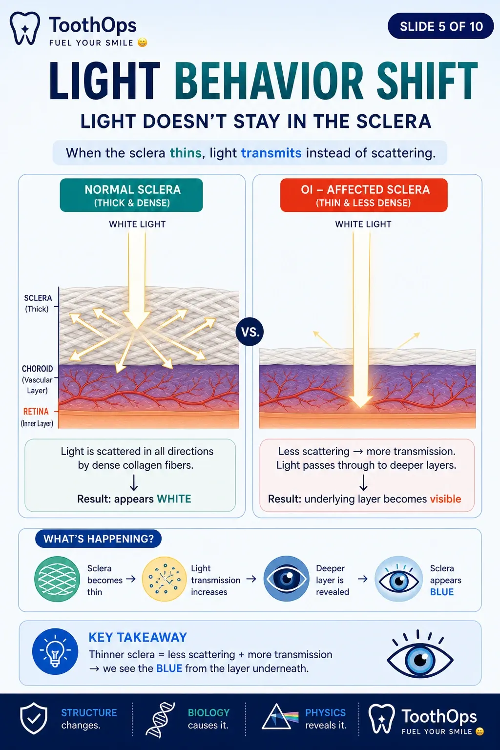
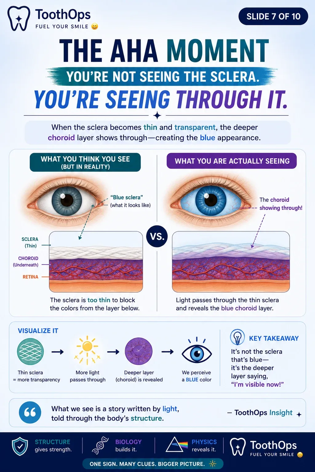
When the sclera thins:
- less light is scattered
- more light passes through deeper layers
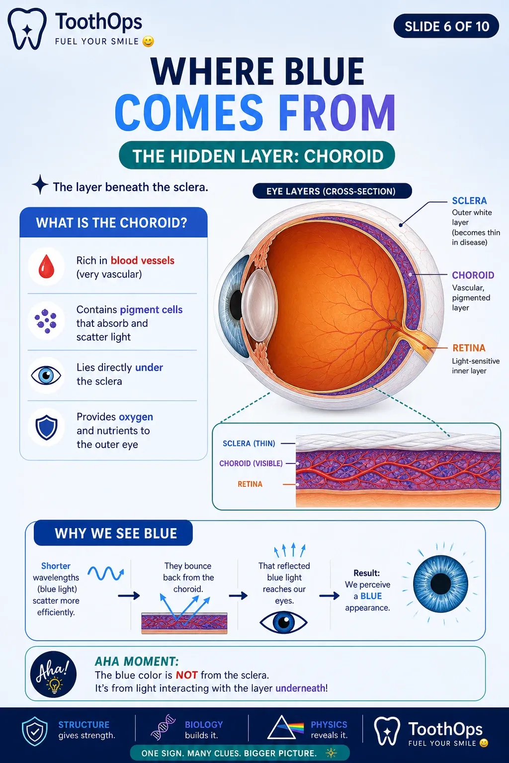
🔬 The Choroid (Hidden Layer)
Underneath the sclera lies the choroid:
- highly vascular tissue
- rich in blood vessels and pigment
As the sclera becomes more translucent:
👉 underlying choroidal tissue becomes visible
🧠 Why the Eye Looks Blue
Shorter wavelengths of light (blue wavelengths):
- scatter more efficiently
- reflect back more prominently through thin tissue
🧠 AHA MOMENT
The sclera itself is not turning blue.👉 You are seeing deeper structures through weakened collagen.
🧠 Signature Framework — Structure → Light → Perception
| Structural Change | Optical Effect | What We See |
|---|---|---|
| Thin collagen | less scattering | more light transmission |
| Increased transparency | choroid visible | blue appearance |
| Collagen defect | altered tissue mechanics | systemic connective tissue clue |
📊 Pattern Recognition Table
| Clinical Finding | Mechanism | Clinical Meaning |
|---|---|---|
| Blue sclera | thin translucent collagen | connective tissue disorder |
| Frequent fractures | weak osteoid matrix | bone fragility |
| Dentinogenesis imperfecta | defective dentin scaffold | tooth fragility |
| Hearing loss | ossicle collagen weakness | systemic involvement |
🧠 Clinical Thinking
When these findings occur together:
- blue sclera
- fractures
- dentin abnormalities
- hearing loss
👉 think:
systemic Type I collagen disorder
⚠️ Critical Exceptions (High-Yield Nuance)
❗ Blue sclera is NOT exclusive to OI
Can also occur in:
- Ehlers-Danlos syndrome
- severe iron deficiency anemia
- connective tissue disorders
- some Marfan-related conditions
❗ Not all OI patients have blue sclera
Severity varies depending on:
- collagen quantity vs quality
- subtype of mutation
- tissue involvement
🧠 Clinical Insight
Blue sclera is a clue—not the diagnosis itself.
🧠 Diagnostic Thinking Framework
Step 1 — Recognize the visual sign
👉 blue sclera
Step 2 — Ask WHY the color changed
👉 altered tissue structure
Step 3 — Connect structure to collagen biology
👉 connective tissue weakness
Step 4 — Look for associated systems
- bones
- teeth
- hearing
- growth
Step 5 — Build the whole-body pattern
👉 Osteogenesis Imperfecta becomes likely
🔥 Key Clinical Insight
A single visual clue can reveal an entire systemic disease process.
🦷 Dentistry Relevance
Dentists may be among the first clinicians to notice:
- dentinogenesis imperfecta
- abnormal tooth translucency
- enamel shearing
- repeated fractures during history-taking
When combined with blue sclera:
👉 these findings may help identify a previously undiagnosed connective tissue disorder.
🧠 Clinical Insight
Teeth and eyes together can sometimes reveal what imaging and laboratory tests have not yet confirmed.
💬 Chairside Translation (Patient-Friendly)
When patients ask:
“Why do my eyes look blue?”
A calm explanation can build enormous trust.
“The white part of the eye is made from strong connective tissue called collagen. In your case, that layer is thinner than usual, so light passes through differently and reflects the deeper layer underneath, which creates the blue appearance. It’s actually an important clue that helps us understand how the connective tissues in the body are built.”
🧠 ToothOps Insight
What we see is not always the structure itself—👉 it is often how light interacts with that structure.
📦 If You Only Remember One Thing
👉 Blue sclera = thin collagen → increased light transmission → deeper vascular tissue becomes visible
✨ Final Takeaway
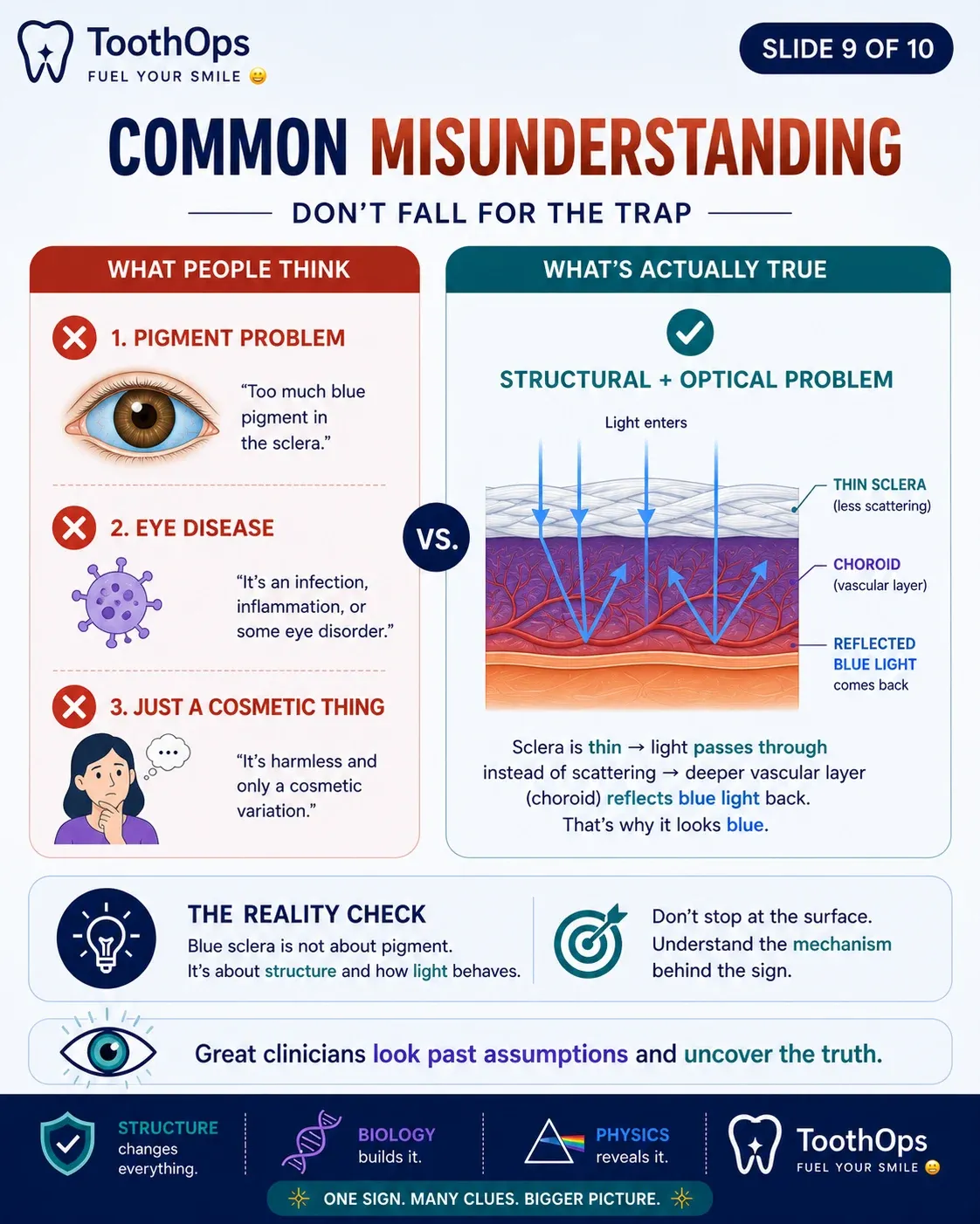
Blue sclera is not a cosmetic feature or random color change.
It is:
- a structural clue
- an optical consequence of collagen failure
- and part of a larger whole-body pattern
Understanding why it happens transforms the finding from something to notice…
👉 into something meaningful enough to change diagnosis, treatment, and patient care.
🧠 Question
A pediatric patient presents with:
- blue sclera
- multiple fractures
- translucent teeth
Which mechanism BEST explains the blue sclera?
A.
Increased melanin deposition in the sclera
B.
Reduced collagen thickness allowing visualization of underlying choroidal tissue
C.
Increased vascular proliferation within the sclera
D.
Calcium deposition altering light reflection
✅ Correct Answer: B
🧠 Explanation
Why B is correct:
- collagen defect → thin sclera
- increased light transmission
- deeper vascular layer becomes visible
Why the others are incorrect:
A.
Pigmentation does not explain the optical transparency mechanism.
C.
The blood vessels are not increased—they become more visible because the sclera is thinner.
D.
Calcium deposition would increase opacity, not transparency.
🧠 High-Yield Learning Point
Blue sclera is fundamentally a problem of transparency and light transmission—not pigmentation.
🧠 Final ToothOps Insight
The body rarely fails in isolation.When structure changes at the molecular level, the effects can appear everywhere—from bone, to teeth, to the way light moves through the eye.
Blue sclera is not simply an eye finding.
👉 It is a visible reminder that:
- biology is interconnected
- structure determines function
- and careful observation can reveal hidden disease long before labs or imaging confirm it.
The best clinicians do not just memorize signs.
👉 They learn to ask:
“What mechanism could connect all of these findings together?”
That is where true clinical reasoning begins.
💬 Final Communication Insight
Patients do not expect perfection. They want clarity, understanding, and confidence that someone truly sees the bigger picture.
@ToothOps | Fuel Your Smile 😊
Stay tuned for more insights and educational content in our blog.
Disclaimer: Content is for educational purposes only and not a substitute for medical or dental care.
© 2025 ToothOps | All Rights Reserved.