
🦷 When the Liver Isn’t Failing — But Still Not Working
 ToothOps5 min read
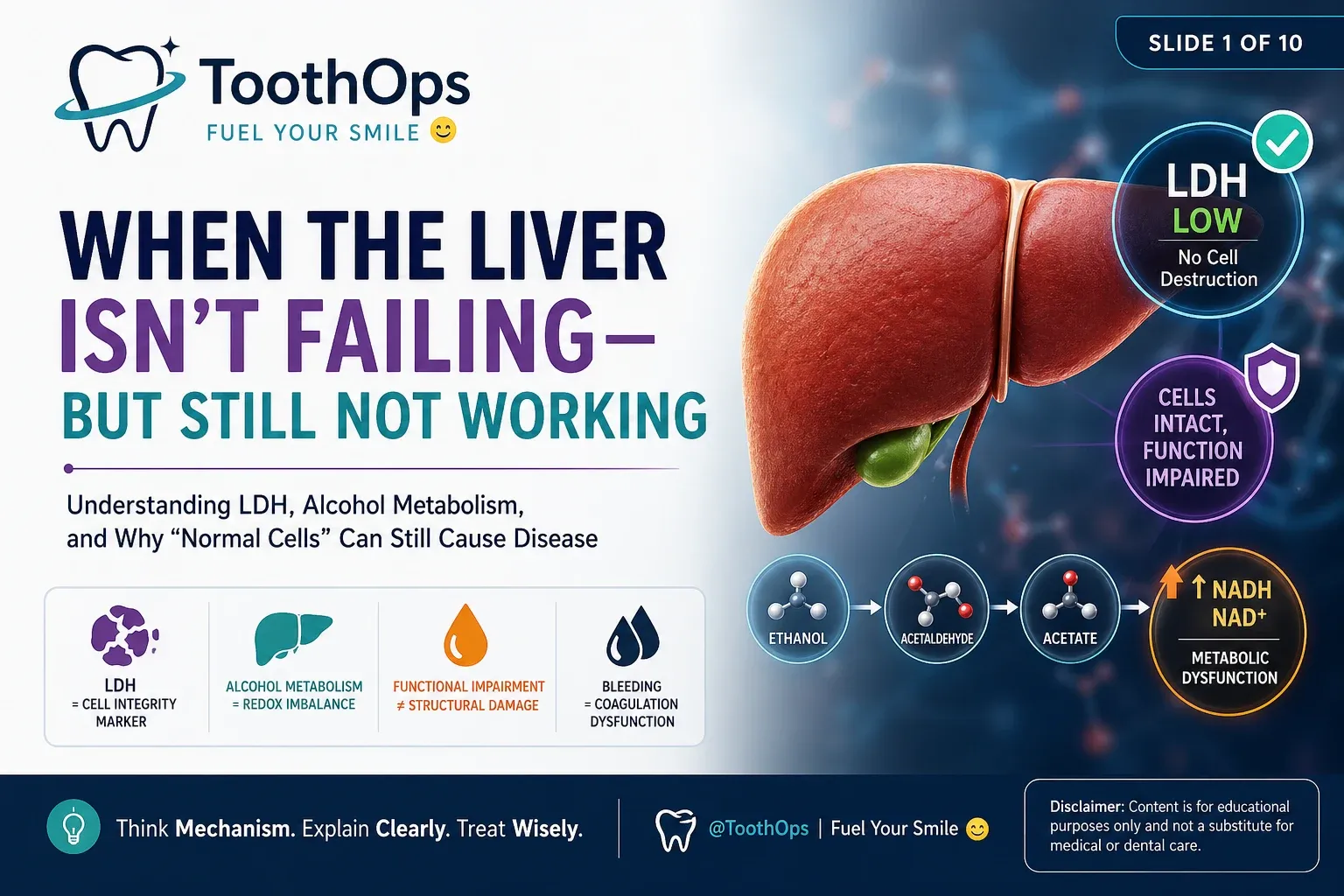
🧠 Understanding LDH, Alcohol Metabolism, and Why “Normal Cells” Can Still Cause Disease
🔍 Introduction — The Misleading Lab Value
In medicine, we’re trained to look for what is elevated.
High enzymes.High markers.High signals of damage.
But sometimes, the most important finding is the opposite:
👉 A value that is low — when you expect it to be high.
That’s exactly what happens with lactate dehydrogenase (LDH).
In this case, LDH isn’t elevated.
It’s low.
And that single detail completely changes how we understand the disease.
🧠 The Big Shift: Damage vs Dysfunction
Most people assume:
👉 If an organ isn’t working → it must be damaged
But biologically, that’s not always true.
There are two fundamentally different states:
⚙️ 1. Structural Damage (Cell Death)
- Cells rupture
- Membranes break
- Intracellular enzymes leak out
👉 This is where LDH rises
⚙️ 2. Functional Impairment (Cells Alive, But Struggling)
- Cells remain intact
- Metabolism is disrupted
- Output is impaired
👉 LDH stays normal or low
📌 This distinction is the core of this entire case:
The liver is not being destroyed.It is metabolically overwhelmed.
🧬 What LDH Actually Measures (Beyond Memorization)
LDH is often taught as a “tissue damage marker.”
That’s incomplete.
🔬 True Mechanism:
LDH is a cytosolic enzyme that catalyzes:
- Pyruvate ⇄ Lactate
- NADH ⇄ NAD⁺
This reaction is essential for:
- Maintaining redox balance
- Allowing glycolysis to continue, especially in low oxygen states
⚠️ Critical Clinical Insight:
LDH is inside the cell.
It only appears in blood when:
👉 The cell membrane is disrupted
📊 Therefore:
| LDH Level | Meaning |
|---|---|
| 🔼 High | Cell death (hemolysis, necrosis, ischemia) |
| 🔽 Normal / Low | Cells intact → no cytolysis |
👉 LDH does NOT measure:
- Inflammation
- Metabolic stress
- Organ function
It measures cellular integrity
🍷 Alcohol Metabolism — Where the Real Problem Begins
At first glance, alcohol seems like a toxin that “damages the liver.”
But the early story is more subtle.
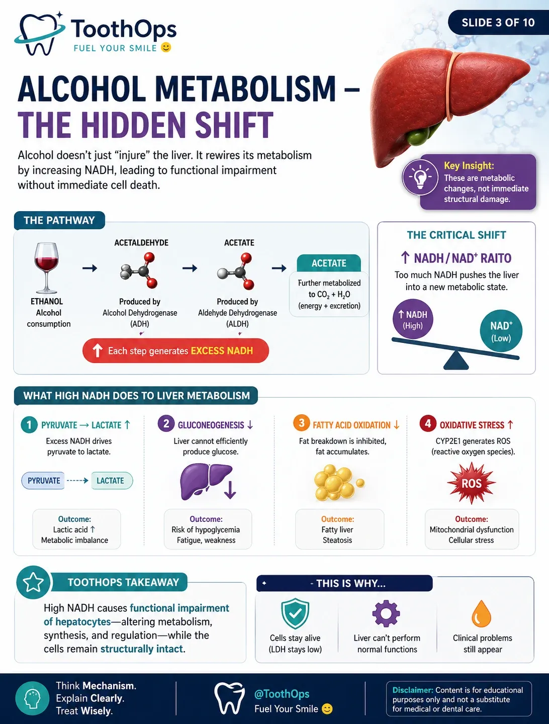
🔬 Step 1: Ethanol Breakdown
Ethanol → Acetaldehyde → Acetate
Enzymes involved:
- Alcohol dehydrogenase (ADH)
- Aldehyde dehydrogenase (ALDH)
- CYP2E1 (MEOS system)
⚠️ Step 2: The Hidden Consequence
These reactions produce large amounts of:
👉 NADH
🔥 Result:
NADH/NAD⁺ ratio increases dramatically
This single shift rewires liver metabolism.
🔄 Metabolic Reprogramming (The Part Most People Miss)
When NADH rises, the liver is forced into a new metabolic state:
1. Pyruvate → Lactate ↑
- Drives lactate production
- Can contribute to metabolic imbalance
2. Gluconeogenesis ↓
- Liver can’t produce glucose efficiently
- Energy regulation becomes unstable
3. Fatty Acid Oxidation ↓
- Fat accumulates in hepatocytes
- Leads to fatty liver changes
4. Reactive Oxygen Species ↑
- Especially via CYP2E1
- Causes oxidative stress and mitochondrial dysfunction
📌 Key Insight:
These are functional metabolic changes Not immediate structural damage
🧠 Why LDH Stays Low in Alcohol-Related Dysfunction
Even though the liver is clearly impaired:
- Bilirubin rises
- Albumin drops
- Coagulation worsens
👉 LDH remains low
Why?
Because:
👉 The hepatocytes are still structurally intact
They are:
- Alive
- Stressed
- Dysfunctional
But not yet:
- Ruptured
- Necrotic
🩺 Connecting This to the Patient
Let’s interpret the case like a clinician:
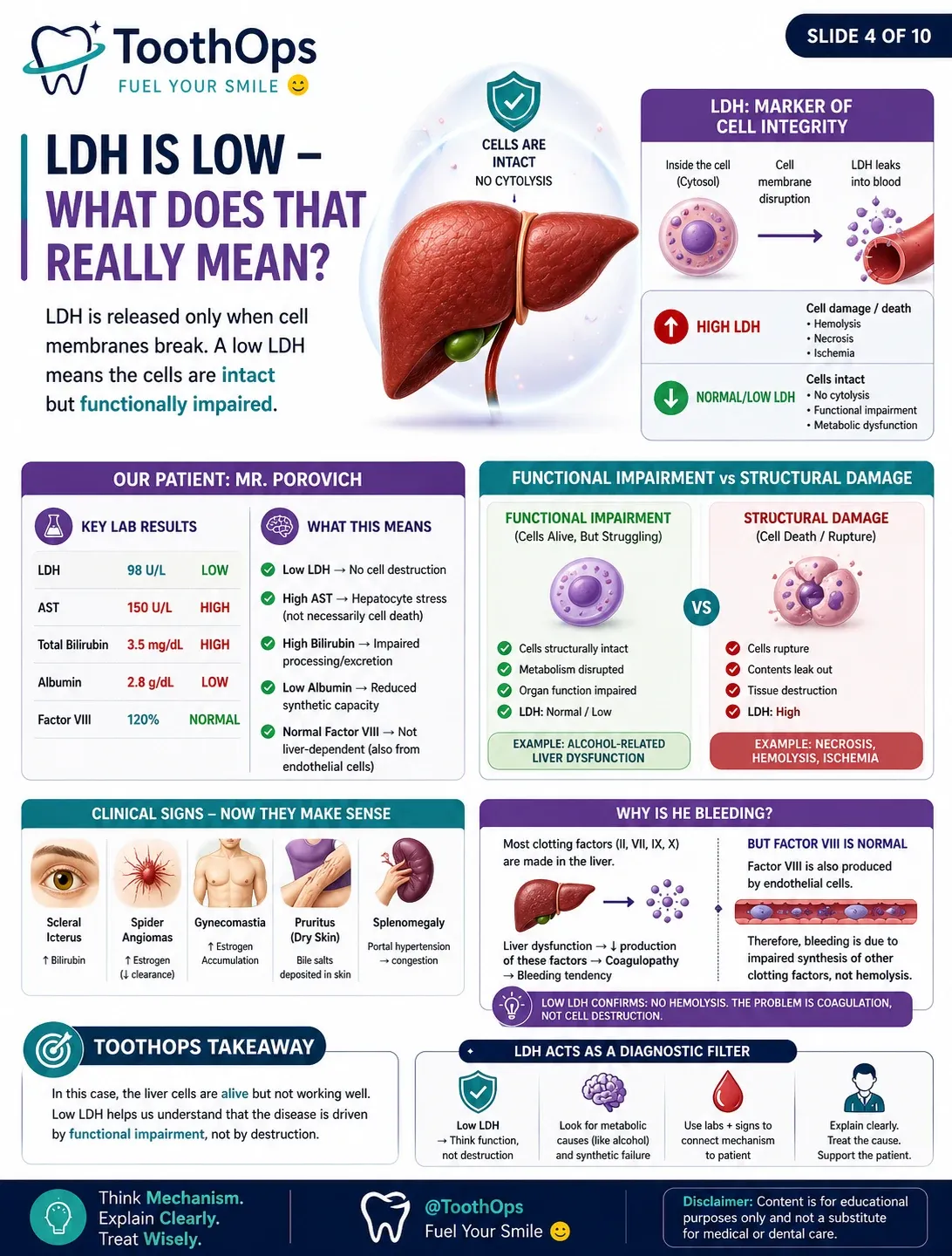
📊 Key Labs:
- LDH ↓ → no cell destruction
- AST ↑ → hepatocyte stress
- Bilirubin ↑ → impaired processing
- Albumin ↓ → decreased synthesis
🧠 Mechanistic Interpretation:
The liver is functionally failingBut not undergoing widespread cell death
🩸 The Bleeding Problem — Explained Correctly
At first, bleeding might suggest:
👉 “Are red blood cells being destroyed?”
But LDH answers that:
❌ No hemolysis
❌ No major cytolysis
So what’s causing the bleeding?
👉 Coagulation dysfunction
🧬 Why?
The liver produces most clotting factors:
- II, VII, IX, X
When liver function declines:
- These factors decrease
- Clotting becomes impaired
⚠️ Important Exception:
Factor VIII remains normal
Why?
👉 It is also produced by endothelial cells
📌 Clinical takeaway:
Bleeding can persist even when Factor VIII is normalBecause the rest of the coagulation system is impaired
🧠 Clinical Signs — Now They Make Sense
Each symptom becomes logical when viewed mechanistically:
- Scleral icterus → bilirubin accumulation
- Spider angiomas → estrogen buildup
- Gynecomastia → impaired hormone metabolism
- Pruritus → bile salt deposition
- Splenomegaly → portal hypertension
⚙️ The Most Important Concept in This Case
🔑 Functional vs Structural Disease
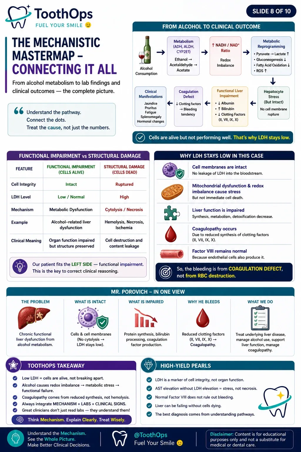
| Feature | Functional Dysfunction | Structural Damage |
|---|---|---|
| Cells | Alive | Dead |
| LDH | Low | High |
| Mechanism | Metabolic failure | Cytolysis |
| Example | Alcohol-related dysfunction | Necrosis / hemolysis |
👉 This patient:
Falls entirely into functional dysfunction
🧠 Why This Matters
Without understanding LDH, you might conclude:
❌ “This is liver cell destruction”
❌ “This is hemolysis”
But with LDH:
👉 You correctly identify:
✅ Chronic metabolic liver dysfunction
✅ Coagulation impairment from synthesis failure
✅ No active cell destruction
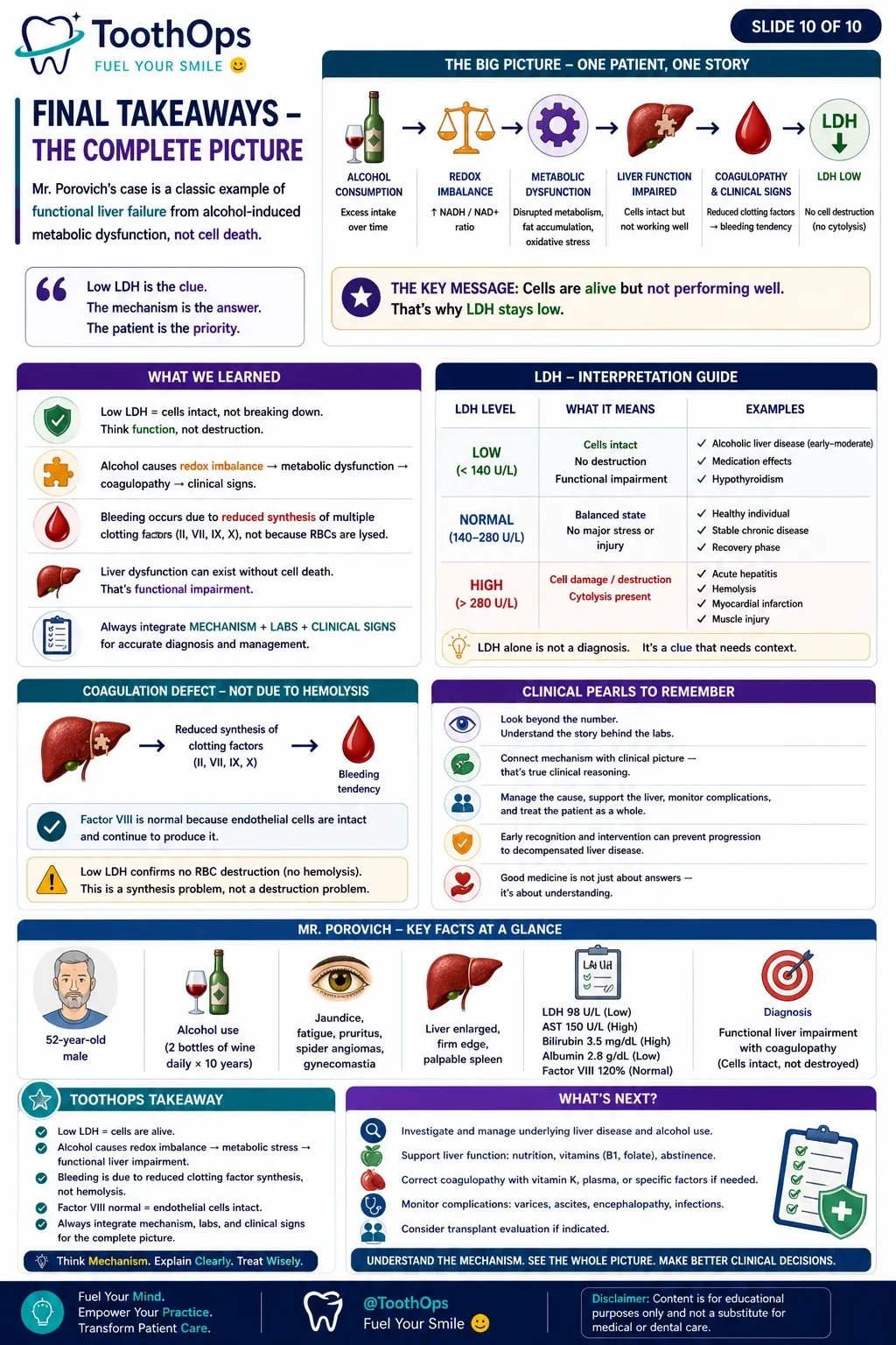
🪶 ToothOps Takeaway
- LDH does not tell you how well cells work
- It tells you whether cells are breaking apart
👉 Low LDH + abnormal liver labs =cells alive, but functionally compromised
🦷 Chairside Explanation
“Your liver cells are still alive, which is good — they’re not breaking apart.But they’re under stress and not working properly right now, which is why we’re seeing issues like bleeding and changes in your lab results.”
🧠 Knowledge Check
Question:
A patient presents with:
- Elevated AST
- Elevated bilirubin
- Low albumin
- Normal LDH
What is the most likely underlying mechanism?
Answer:
👉 Functional hepatocellular impairment without cytolysis
Explanation:
- Low LDH → no cell membrane rupture
- High AST → cellular stress
- Low albumin + high bilirubin → impaired liver function
👉 Therefore:Cells are intact but metabolically dysfunctional
📚 Clinical Relevance Summary
- LDH = cell integrity marker
- Alcohol metabolism = redox-driven metabolic disruption
- Liver disease can exist without cell death
- Coagulopathy = synthetic failure, not RBC destruction
@ToothOps | Fuel Your Smile 😊
Stay tuned for more insights and educational content in our blog.
Disclaimer: Content is for educational purposes only and not a substitute for medical or dental care.
© 2025 ToothOps | All Rights Reserved.