
🦷 Caries Diagnosis 101: How Dentists Really Know When a Spot Is a Cavity (and When It’s Not)
 ToothOps4 min read
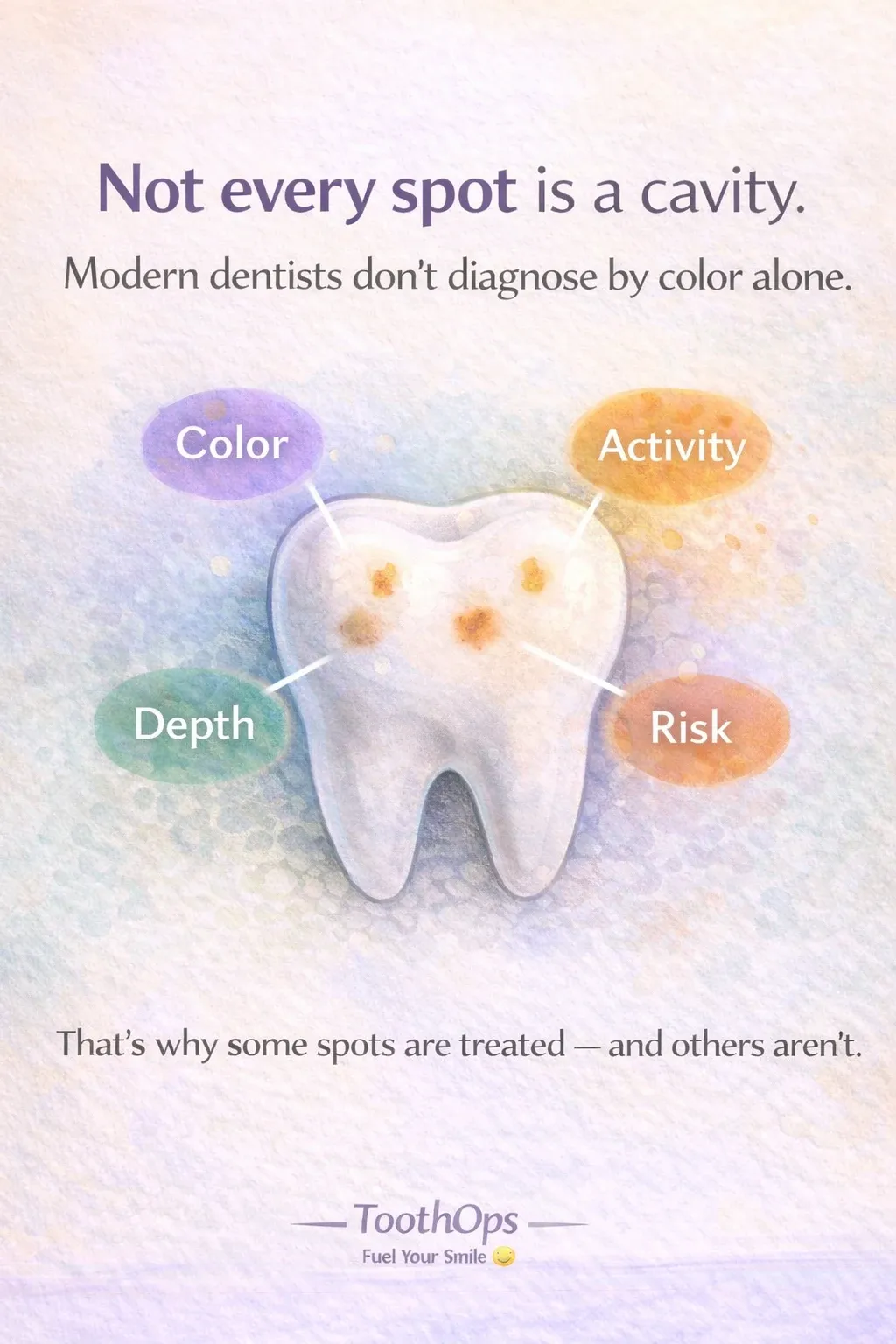
If you’ve ever stared at a tiny white or brown spot on your tooth and thought:
“Is this… a cavity?”
You’re not alone — and here’s the secret:
⭐ Dentists don’t decide based on color alone.
⭐ Not every white or brown spot needs a filling.
⭐ Modern diagnosis is WAY more precise than it used to be.
Today’s dentistry uses a combination of clinical evidence, radiographs, and risk assessment to determine whether a tooth needs remineralization, monitoring, or intervention.
Let’s break down how caries diagnosis actually works — so you can understand your mouth, your radiographs, and your treatment plan like a pro.
1️⃣ The Foundation: Dentistry Doesn’t “Guess” Anymore
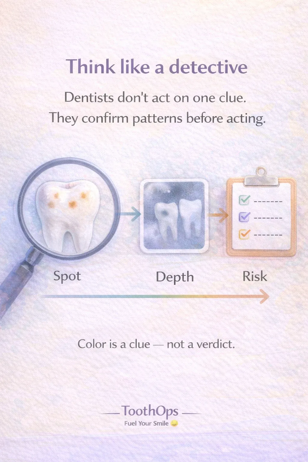
Proper diagnosis requires a structured, step-by-step evaluation of the whole patient — not just the tooth.
Diagnosis integrates the following essential steps:
- Chief concern
- Medical history
- Dental history
- Clinical soft tissue exam
- Tooth-by-tooth evaluation
- Radiographs
- Caries risk assessment
- Treatment planning
🧠 Analogy: Think of it like a detective show. The dentist isn’t judging based on “clues” (color, shadows) — they gather evidencefrom multiple sources before reaching a verdict.
2️⃣ What Caries Actually Looks Like
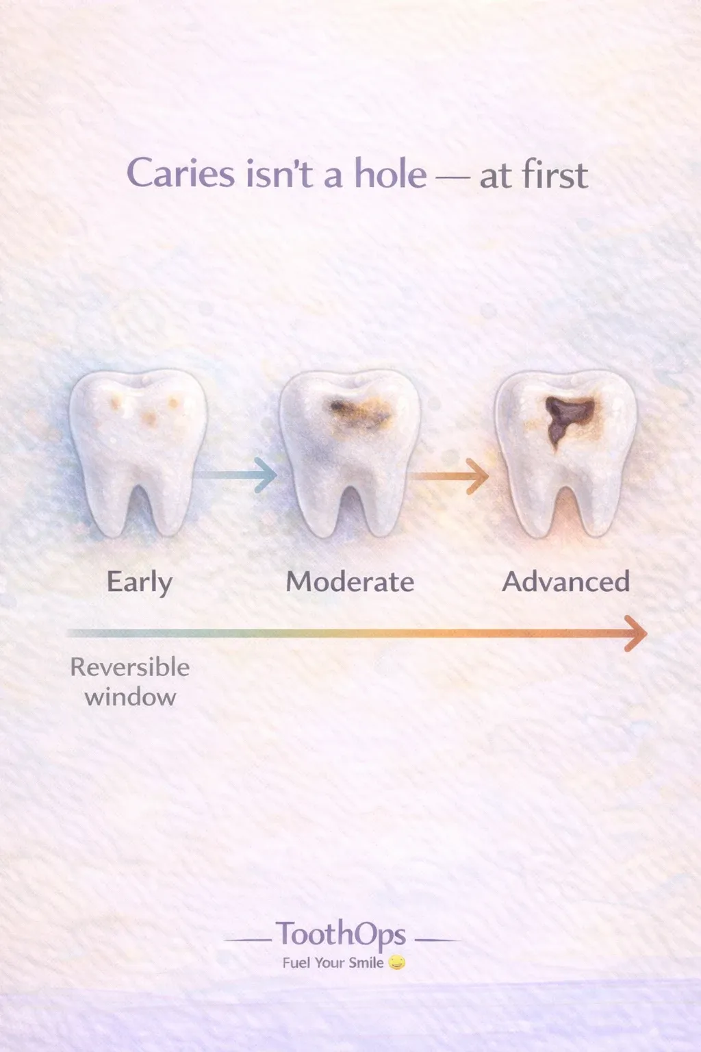
Caries isn’t a hole — not at first.
Initial Stage / White Spot Lesion
- Chalky
- Matte
- Rough
- Surface intact
- Reversible with remineralization
Moderate Stage
- Possible microcavitation
- Shadowing
- Opacity changes
- May need minimally invasive treatment
Advanced Stage
- Actual cavitation
- Dentin involvement
- Likely needs restoration
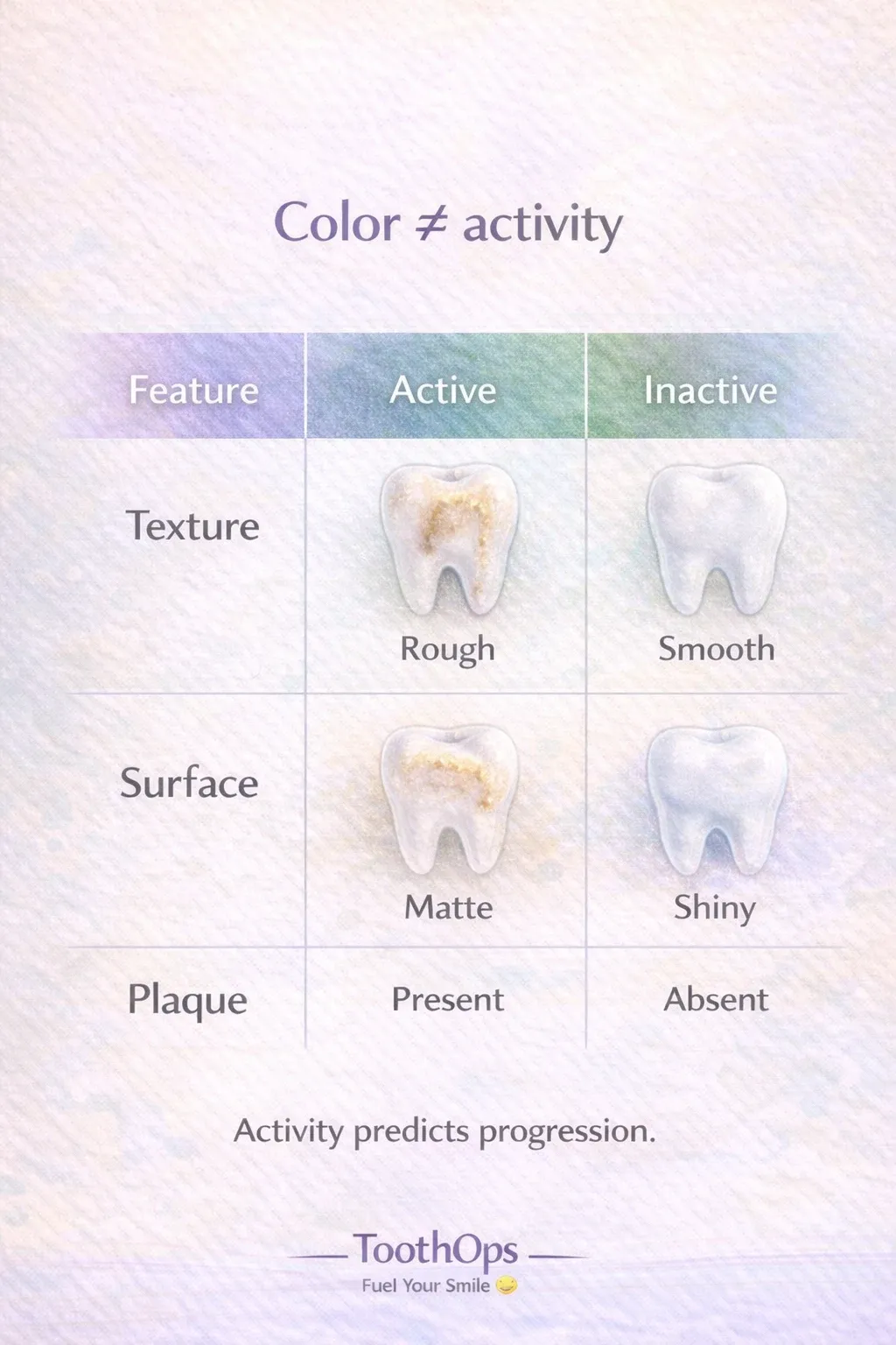
🧠 Quick checklist for dental students:Color ≠ activityRoughness + plaque = activityShiny + hard = inactive
3️⃣ Radiographs: The “X-Ray Vision” Behind Diagnosis
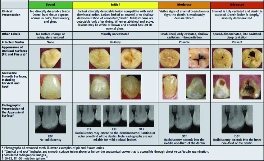
Radiographs aren’t meant to “look cool” on the lightbox — they’re a diagnostic map.
The radiographic stages:
- E0 – Sound enamel
- E1 – Outer ½ of enamel
- E2 – Inner ½ of enamel
- D1 – Outer ⅓ of dentin
- D2 – Middle ⅓ of dentin
- D3 – Inner ⅓ of dentin, near pulp
🧠 **Analogy:**Think of enamel like the “outer wall” of a castle.If the damage is only on the outer stones (E1/E2), you can repair it.If it’s into the foundation (D2/D3), you need structural reinforcement (a restoration).
4️⃣ Lesion Activity: The Step Students AND Patients Miss
In Chapter 2, activity assessment is one of the biggest indicators of whether a lesion needs treatment. Chapter_2_Dental_Caries__Etiolo…
Active Lesions (Disease progressing):
- Chalky, matte
- Rough texture
- Soft dentin
- Found in plaque-retentive areas
- Often near gingiva
Inactive Lesions (Disease arrested):
- Shiny
- Smooth
- Hard to explorer/light pressure
- No plaque stagnation
💡 **Pro Tip (for everyone):**An inactive lesion can stay “scarred” for decades without needing a filling.Dentists only intervene when the disease is progressing, not just visible.
5️⃣ Caries Risk Assessment: Diagnosis ≠ Treatment Plan
This is where so many patients — and new dental students — get confused.
Diagnosis tells us:👉 What is happening on each tooth.
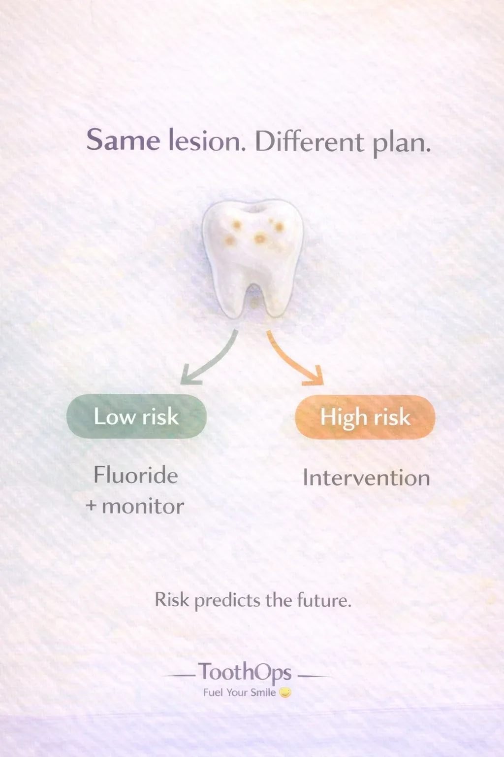
Risk assessment tells us:👉 What’s likely to happen next.
CAMBRA highlight that risk is based on:
- Diet frequency
- Saliva quantity/quality
- Fluoride exposure
- Past disease (white spot, cavitations, new lesions)
- Socio-behavioral factors
🧠 Analogy: Diagnosis is the weather today. Risk assessment is the forecast.
That forecast determines whether a lesion is:
- Remineralizable
- Monitorable
- Or ready for operative treatment
6️⃣ When Dentists Choose NOT to Drill
A big misconception:“Any spot = filling.”
But modern, evidence-based dentistry avoids unnecessary drilling because:
✔ Tooth structure is precious
✔ Early lesions can heal
✔ Risk-driven management prevents progression
Many E1/E2 lesions are nonoperative in low-risk patients — especially if they’re non-cavitated and inactive.
Drilling treats the hole.Risk management treats the disease.
Both matter — but they’re not the same thing.
7️⃣ Why This Matters for You (Dental Student, Pre-Dental, or Patient)
For Dental Students:
You’ll be tested on:
- Lesion staging
- Activity assessment
- ICDAS–radiographic correlation
- CAMBRA risk factors
- Treatment thresholdsUnderstanding these concepts now makes PBLs, OSCEs, and clinic smoother.
For Pre-Dentals:
Talking about “disease vs lesion” and “activity vs depth” blows interviewers away.It shows you understand dentistry as chronic disease management, not drilling.
For Patients:
This helps you advocate for yourself and understand why your dentist may say:
- “Let’s monitor this.”
- “Let’s remineralize this spot.”
- “Let’s address your snacking or dry mouth first.”
It’s not guessing. It’s science.
💪 ToothOps Takeaway
Caries diagnosis is way more than spotting dark areas.It’s a fine balance of biology, behavior, and evidence-based assessment.
When you understand depth, activity, and risk, you understand exactly why a dentist chooses fluoride, monitoring, or a filling.
Your smile isn’t just a set of teeth — it’s a story your habits, biology, and environment are constantly writing.
@ToothOps | Fuel Your Smile 😊
Stay tuned for more insights and educational content in our blog.
Disclaimer: Content is for educational purposes only and not a substitute for medical or dental care.
© 2025 ToothOps | All Rights Reserved