
🧬 When the Immune System Overreacts: The 4 Types of Hypersensitivity (I–IV) Explained
 ToothOps7 min read
We love the immune system… until it turns a small problem into a big one.
Allergies, autoimmune diseases, serum sickness, contact dermatitis — they look totally different in clinic, but on an exam they all live under one umbrella: hypersensitivity reactions.
In simple terms, hypersensitivity = an exaggerated or misdirected immune response that ends up harming the host.
The good news for your brain (and your PBL notes):There are only four main patterns — Type I, II, III, and IV. Once you understand the logic of each type, the long list of diseases finally stops feeling random.
This ToothOps guide walks you through all 4 types — what they are, how they work, classic examples, and where they matter in dentistry.
1️⃣ The Big Picture: 4 Types on One Page
Before diving into details, anchor the whole map in your head:
| Type | Main Weapon | Timing | Where It Acts | Classic Examples |
|---|---|---|---|---|
| Type I | IgE + mast cells | Immediate(minutes) | Mucosa, skin, vasculature | Anaphylaxis, allergic asthma, allergic rhinitis, some food allergies |
| Type II | IgG or IgM bound to cell surfaces | Hours–1 day | Specific cells or membranes | Goodpasture syndrome, autoimmune hemolytic anemia, immune thrombocytopenia, myasthenia gravis, Graves’ disease |
| Type III | Immune complexes(antigen–antibody) floating in serum | 1–2 days | Vessels, kidneys, joints | Serum sickness, Arthus reaction, SLE, some vasculitides, rheumatoid arthritis |
| Type IV | T-cells (CD4+ and CD8+) | 2–3 days (delayed) | Skin, lungs, organs | TB skin test, contact dermatitis, granulomas (TB, sarcoid), chronic transplant rejection |
🧠 Memory map:
- Types I–III = antibody-driven (humoral)
- Type IV = T-cell–driven (cellular)
Keep that hierarchy in mind as we zoom in.
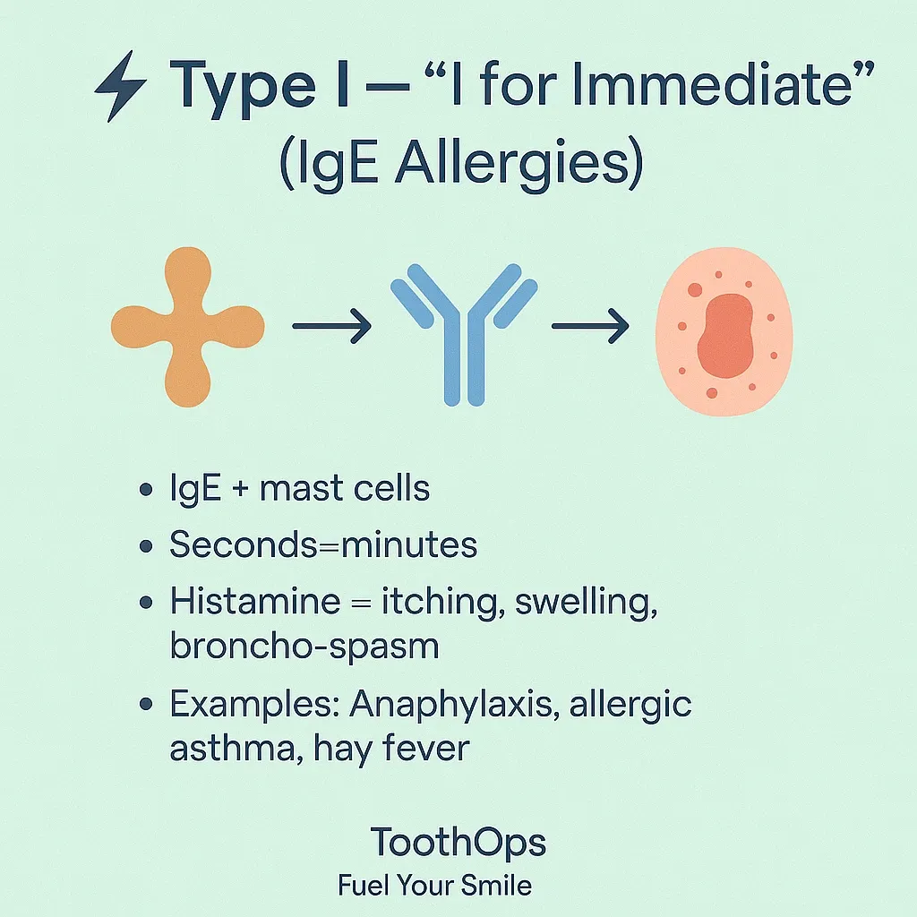
2️⃣ Type I – Immediate Hypersensitivity (“I for Immediate, IgE, Allergy”)
**Core idea:**Your body makes IgE against an otherwise harmless antigen (pollen, dust mites, foods, insect venom). IgE coats mast cells. On re-exposure, the allergen cross-links IgE on those mast cells → they explode with mediators.
Mechanism in two exposures
- First exposure (sensitization):
- Allergen enters the body.
- Antigen-presenting cells show it to T-cells → Th2 response.
- Th2 cells release IL-4/IL-13 → B-cells class switch to IgE.
- IgE binds Fc receptors on mast cells and basophils and just… waits.
- Second exposure (reaction):
- Same allergen returns.
- Cross-links IgE on mast cells.
- Mast cells degranulate:
- Early phase (preformed mediators): histamine, serotonin, eosinophil chemotactic factors
- Late phase (newly synthesized): leukotrienes, prostaglandins, platelet-activating factor
Clinical effects:
- Bronchoconstriction
- Vasodilation and increased vascular permeability → swelling, urticaria
- Increased mucus secretion
- In severe cases: anaphylactic shock
Classic examples:
- Anaphylaxis to foods, drugs, or insect stings
- Allergic asthma (extrinsic)
- Allergic rhinitis (“hay fever”)
- Some acute urticaria reactions
🧠 **Analogy Box:**Think of mast cells as granule-filled balloons primed with IgE tripwires. The first exposure ties the tripwires on. The second exposure pulls them all at once.
💡 **Dental Pro Tip:**Always review:
- Drug allergies (especially penicillins, local anesthetics).
- Latex sensitivity.Have an emergency plan for anaphylaxis (epinephrine, oxygen, airway protocol) — this aligns with ADA and emergency preparedness guidelines.
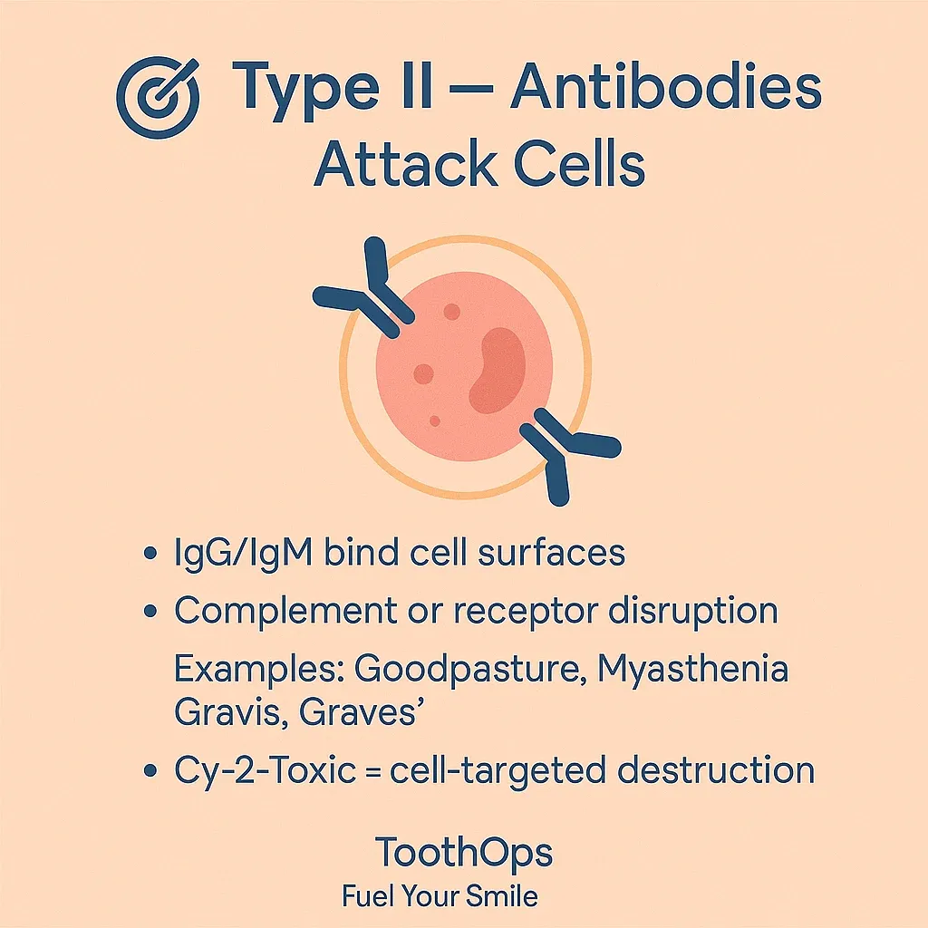
3️⃣ Type II – “Cy-2-Toxic”: Antibodies Against Cell Surfaces
**Core idea:**In Type II, antibodies attach directly to cells or extracellular structures and mark them for destruction or interfere with their receptors.
Main antibodies: IgG, IgMMain mechanisms:
- Complement-mediated lysis & opsonization
- IgG/IgM bind a cell surface antigen.
- Classical complement pathway activates → membrane attack complex (MAC) & opsonins.
- Result: cell lysis and phagocytosis.
- Antibody-dependent cell-mediated cytotoxicity (ADCC)
- Antibody-coated cells are killed by NK cells, macrophages, or neutrophils.
- Receptor interference
- Antibody blocks receptor → loss of function.
- Antibody stimulates receptor → overactivity (rare but high yield).
Classic examples to anchor:
- Goodpasture syndrome
- Antibodies against glomerular and alveolar basement membranes → hematuria + hemoptysis.
- Autoimmune hemolytic anemia
- Antibodies against RBCs → hemolysis.
- Immune thrombocytopenia
- Platelets targeted → low platelet count, bleeding.
- Myasthenia gravis
- Antibodies block acetylcholine receptors at the neuromuscular junction → fatigable weakness.
- Graves’ disease
- Antibodies stimulate TSH receptors → hyperthyroidism.
🧠 **Analogy Box:**Type II is like sticking a “target” sticker on a specific cell. The immune system then attacks that tagged cell specifically.
💡 **Dental Pro Tip:**Patients with Type II processes may present with:
- Anemia (fatigue, pallor, tachycardia)
- Thrombocytopenia (easy bruising, petechiae)Screen for bleeding risk before extractions or surgeries, and coordinate with the patient’s physician about platelet counts and medication adjustments.
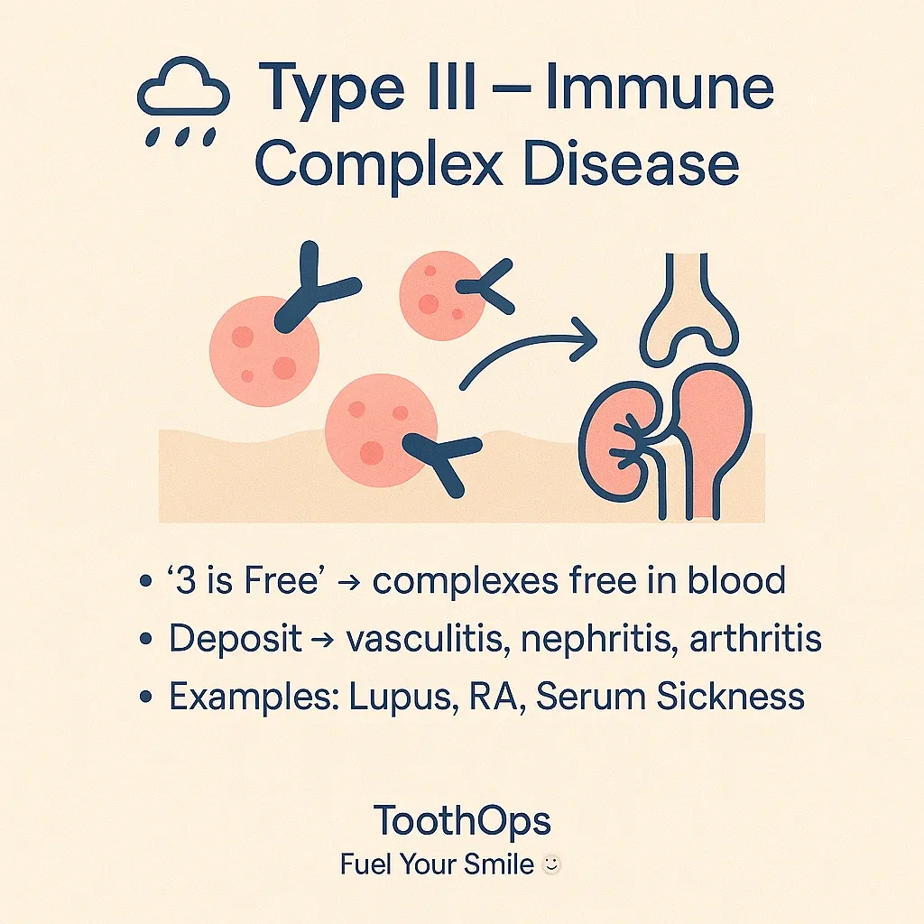
4️⃣ Type III – “3 Is Free”: Immune Complex Disease
**Core idea:**Antigen–antibody complexes form in the circulation and are not cleared efficiently. They float freely (“3 is free”) until they deposit in tissues, triggering inflammation.
Where complexes land most often:
- Blood vessels → vasculitis
- Kidneys → immune complex glomerulonephritis
- Joints → arthritis
These deposited complexes activate complement → C3a, C5a → neutrophil recruitment → tissue damage.
Classic generalized reactions:
- Systemic lupus erythematosus (SLE) – multiple organ immune-complex deposition.
- Rheumatoid arthritis (RA) – immune complex-mediated joint inflammation.
- Serum sickness
- Occurs 1–2 weeks after exposure to foreign proteins/drugs.
- Features: fever, rash, arthralgia, lymphadenopathy, proteinuria.
Classic localized reaction:
- Arthus reaction
- Local, subacute skin reaction at the injection site in a previously sensitized person.
- Painful, red, and indurated area hours after vaccination or injection.
🧠 **Analogy Box:**Think of immune complexes as sticky glitter in the bloodstream. Once it settles in places like vessel walls or glomeruli, the cleanup response causes collateral damage.
💡 **Dental Pro Tip:**Patients with SLE or RA often:
- Take steroids or immunosuppressants (increased infection risk, delayed healing).
- Have TMJ or joint involvement affecting comfort in the chair.Gentle technique, shorter appointments, and close communication with their medical team elevate your standard of care.
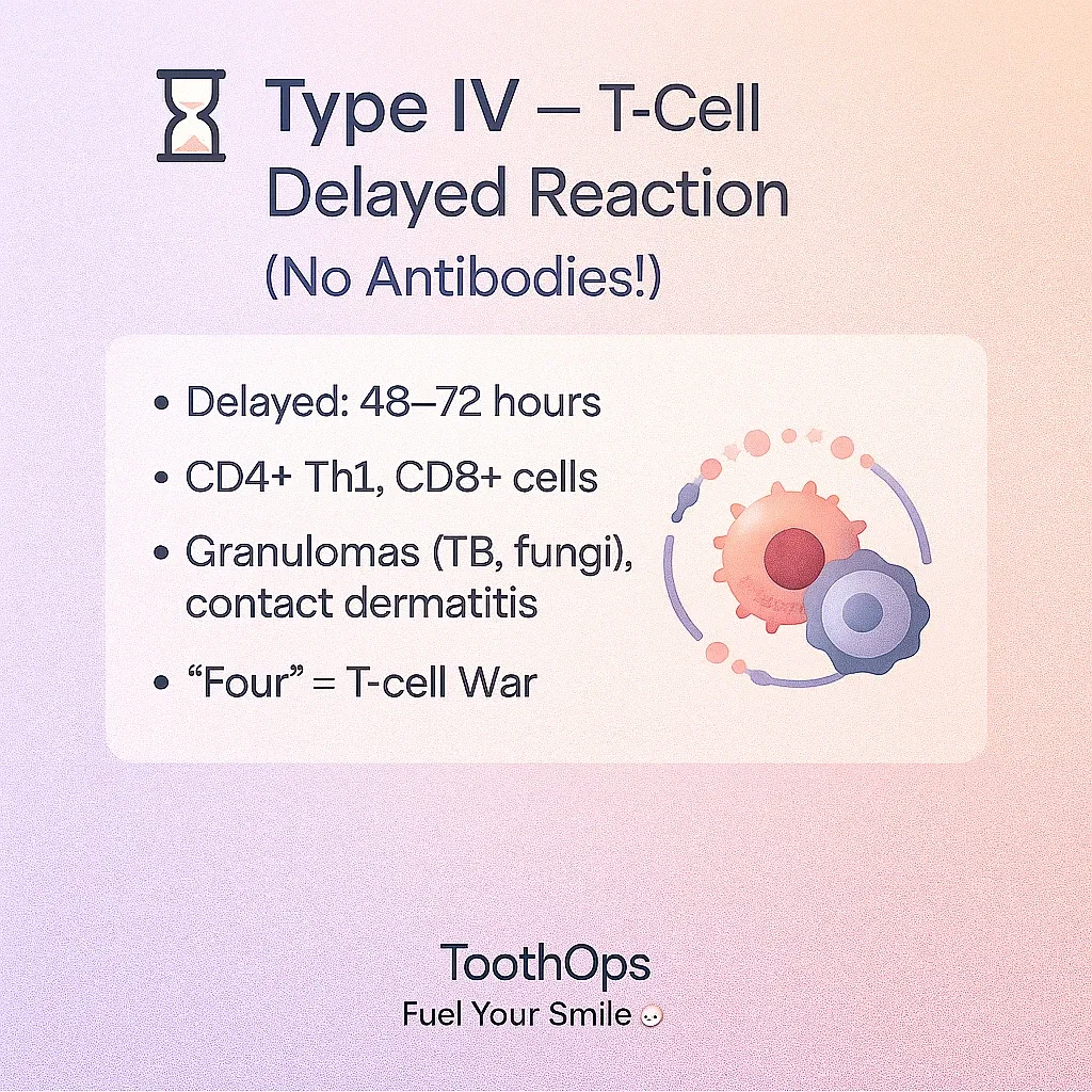
5️⃣ Type IV – Delayed, T-Cell–Mediated Hypersensitivity
**Core idea:**Type IV reactions are driven by **T-lymphocytes, not antibodies.**They are delayed (48–72 hours) because T-cell activation and recruitment take time.
Key players:
- CD4+ Th1 cells – secrete cytokines (especially IFN-γ).
- CD8+ cytotoxic T-cells – directly kill infected/altered cells.
- Macrophages – activated by IFN-γ, participate in granuloma formation.
Two major patterns
- Direct cytotoxicity
- CD8+ T-cells recognize antigen on cells and kill them.
- Delayed-type hypersensitivity (DTH) / granuloma formation
- Antigen-presenting cells (macrophages) present antigen to CD4+ T-cells.
- Th1 cells secrete IFN-γ → activates macrophages.
- If organism is hard to clear (TB, fungi), macrophages and T-cells wall it off → granuloma.
Classic examples:
- Tuberculin skin test (PPD)
- Induration at 48–72 hours = memory T-cell–mediated response.
- Contact dermatitis
- Nickel jewelry, poison ivy, some cosmetics or gloves → itchy, eczematous rash exactly where allergen touched the skin.
- Granulomatous diseases
- TB, histoplasmosis, some fungal infections, sarcoidosis (non-caseating granulomas).
- Chronic transplant rejection & graft-versus-host disease
🧠 **Analogy Box:**Type IV is like a special-operations unit: slower to mobilize, but highly
targeted. If they can’t kill the invader, they build a “prison wall” (granuloma) around it.
💡 **Dental Pro Tip:**Think Type IV when a patient says:
- “I only react days later to that metal or cosmetic.”
- “This rash appears exactly where my watch/denture/material touches.”
For dental materials (metals, resins, flavors), consider patch testing via dermatology/allergy and collaborate on alternative materials.
6️⃣
Comparing All 4 Types: High-Yield Exam & Clinic Snapshot
Use this quick checklist when you’re trying to classify a disease:
- Is IgE involved? Is it immediate?→ Type I (anaphylaxis, allergic asthma, rhinitis).
- Are antibodies binding directly to cell surfaces or receptors?→ Type II (Goodpasture, AIHA, ITP, myasthenia, Graves’).
- Are immune complexes floating in serum and depositing in tissues?→ Type III (SLE, RA, serum sickness, Arthus reaction).
- Are T-cells and macrophages the main players, delayed by days?→ Type IV (TB test, contact dermatitis, granulomas, chronic rejection).
🧠 Exam Strategy Box:
- “Immediate, IgE, allergies, anaphylaxis” → Type I
- “Cytotoxic, cell surface, complement, receptors” → Type II
- “Immune complexes, vasculitis, nephritis, arthritis” → Type III
- “Delayed, T-cells, granulomas, contact dermatitis” → Type IV
a question stem mentions serum sickness, Arthus, or immune complex vasculitis, your brain should instantly flash: Type III.If it mentions PPD, poison ivy, or nickel rash days later, think Type IV.
7️⃣ ToothOps Takeaway: From Memorizing to Understanding
Hypersensitivity doesn’t have to be a wall of memorized lists.
When you understand what weapon the immune system is using and where it’s misfiring, the patterns become logical — and suddenly those PBL cases, NBDE-style questions, and OSCE scenarios feel a lot more manageable.
Small action you can take today:
Pick one disease from each type and explain it out loud to yourself or a friend in two minutes.If you can teach it simply, you own it.
You’re not just memorizing immunology; you’re building the mental framework you’ll use as a future clinician to keep real patients safe.
@ToothOps | Fuel Your Smile 😊
Stay tuned for more insights and educational content in our blog.
Disclaimer: Content is for educational purposes only and not a substitute for medical or dental care.
© 2025 ToothOps | All Rights Reserved